Osteogenesis: How Does Bone Growth Occur?
Osteogenesis is the biological process of bone formation and growth. It begins in the eighth week of embryonic development and is also the process on which the repair of fractures depends. It should be borne in mind that the bones of the human skeleton are derived from three embryonic structures:
- The mesoderm.
- The somites.
- The neural crest.
This process of osteogenesis, also called ossification, consists of the transformation of pre-existing tissue into bone tissue. To carry out this transformation there are two mechanisms:
- Endochondral ossification : it is a more complex process that takes place in two stages. First, the initial tissue is replaced by cartilage and later it ossifies.
- Intramembranous ossification : this is the direct transformation of the original tissue by bone tissue.
Physiology of bone formation and growth
Embryonic structures
The mesoderm is one of the three cell layers from which the entire embryo develops. Initially, there are three layers of cells: an outer, an intermediate, and an inner. The mesoderm is the middle layer and gives rise to the bones of the extremities.
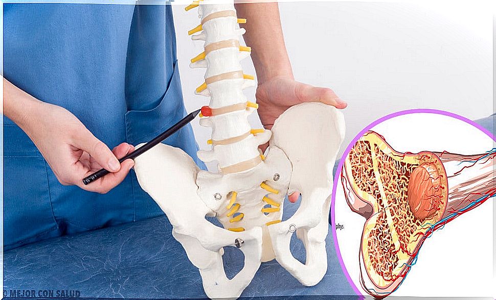
Somites are transient embryonic structures fundamental for the development of the pattern of segmented structures typical of vertebrates. Moreover, from the somites derive the bones that are part of the central axis of the body or axial skeleton. These are the hyoid, ribs, spine, sternum, and skull and hearing bones.
Finally, the neural crest is a transitory cell formation, typical of the first stages of development. Its fundamental characteristic is the pluripotentiality of its cells.
In other words, the cells of the neural crest can give rise to almost any type of definitive structure of the body. For example, they give rise to the craniofacial bones and cartilage.
Endochondral ossification
The endochondral ossification process is characterized by the formation of cartilage from embryonic tissue and its subsequent ossification, which leads to bone growth. We can divide this process into 5 stages:
- First, cells express two transcription factors: PAX 1 and Scleraxix, which are essential for the activation of genes that will turn the original cells into cartilage. This is possible thanks to the fact that cells induce neighboring cells to do so in a paracrine manner.
- The cells that have expressed the necessary genes then clump together and become chondrocytes.
- These chondrocytes multiply very quickly, thus forming a kind of “template” for the future bone.
- Subsequently, the multiplication stops and the chondrocytes increase in size. Likewise, these chondrocytes modify the components they release for the formation of the matrix that can be calcified thanks to calcium carbonate.
- Finally, the cartilage mold is traversed by the forming blood vessels. Chondrocytes die progressively, being replaced by osteoblasts.
Osteoclasts are cells responsible for bone destruction whose activity must always be kept in balance with the activity of osteoblasts. This is because the proper formation and growth of bones depends on this balance.
Intramembranous ossification
Through the intramembranous ossification process, the flat bones of the skull form and grow. This ossification occurs within a connective tissue membrane.
Of the cells in this membrane, some will become osteoblasts, the cells that form the bone matrix. While others become cells that are part of the small blood vessels that supply the bones.
The osteoblasts group together forming what is known as the ossification center, around which the bone will progressively form. This is because these cells synthesize and secrete components that are necessary to create a matrix capable of capturing calcium salts.
What Happens When Osteogenesis Fails?
Imperfect osteogenesis
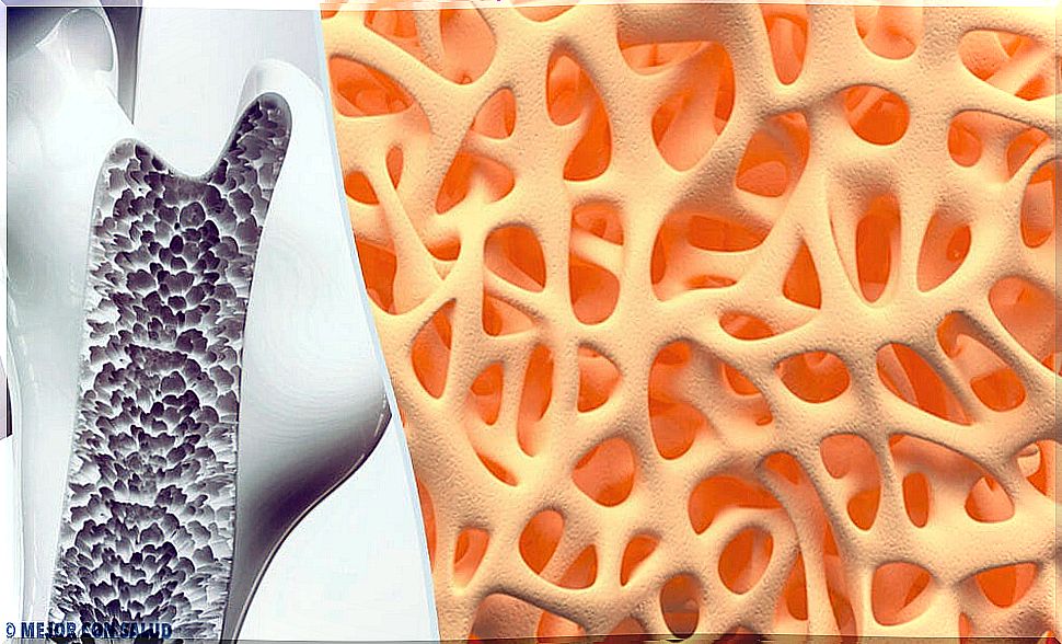
If the process of osteogenesis is not carried out correctly, we find osteogenesis imperfecta, also known as crystal bone disease, it is a genetic disease, characterized by excessive brittleness of the bones.
People who suffer from it constantly suffer fractures in addition to other symptoms. For example: imperfect dentinogenesis or the presence of blue sclera.
The disease occurs as a consequence of mutations that alternate collagen, an essential component in the bone matrix. The lack of this substance is responsible for the excessive fragility of the affected bones.
It is a rare disease due to the small number of people affected. It has a highly variable prognosis, since there are different types of severity. In addition, the evolution of the disease itself also varies greatly between patients and depends on a large number of factors.
There is no specific method of diagnosis. The disease is usually diagnosed through medical history and physical examinations. It could be detected more specifically with a collagen analysis of the skin or with genetic methods.
There is also no treatment, other than the control of symptoms. Specialists recommend physical exercise, physical therapy, use of pain relievers and, in some cases, surgery.