Treatments Of Hilum And Great Vessels
Hilar and great vessel injuries are challenging for surgeons, requiring complex surgical procedures and maneuvers.
Injuries to the hilum and great vessels are usually caused by gunshot or sharps attacks, car accidents, crushing, or errors or difficulties that occur during medical procedures.
Just a few decades ago, the death rate from injuries to the hilum and great thoracic vessels was as high as 62%. Today the range is between 4% and 7%. Still, they are still considered one of the most complex injuries in trauma.
On the other hand, abdominal vascular injuries are those with the highest levels of mortality and morbidity, among all the injuries that someone with severe trauma may suffer. These lesions are often multiorgan.
The great vessels and the hilum

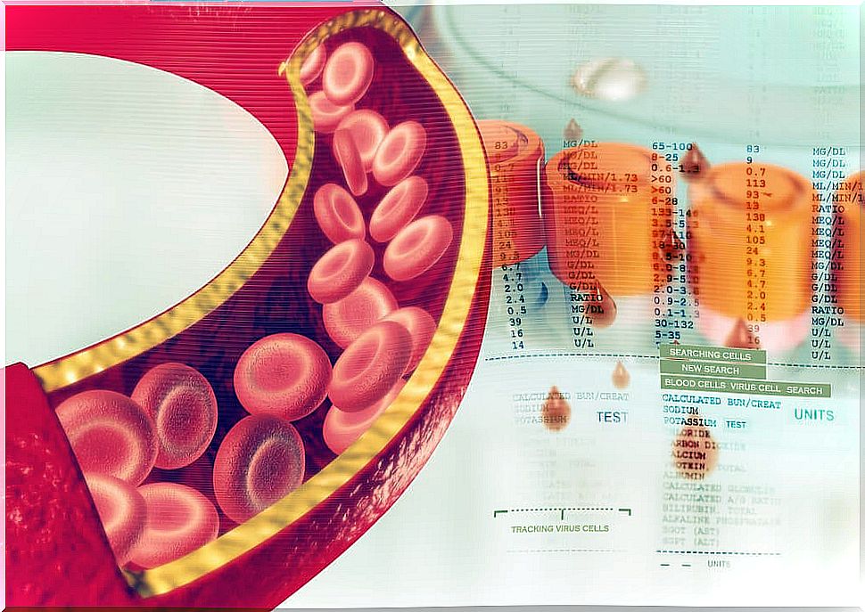
The large veins and arteries that enter and exit the heart are called great vessels . They are basically made up of the aorta, the superior and inferior vena cavae, and the pulmonary veins.
Within the great vessels are the subclavian, carotid, bronchial, posterior intercostals, middle esophageal and iliac, among many others.
The hilum, for its part, is the name given to the concave part of an organ, when it is the point of entry or exit of blood vessels and other structures. They are the hepatic hilum, the pulmonary hilum, the hilum of a lymph node, the renal hilum, the adrenal gland, the anterior olivary and the spleen.
Types of Hilar and Great Vessel Wounds
Wounds of the hilum and great vessels can be of two types: penetrating or non-penetrating. Penetrating wounds are the most frequent and are characterized by giving rise to an open wound, as there is perforation of the skin.
Non-penetrating injuries are more rare and dangerous. They are produced by crushing. The vessel is compressed against the bone, or a fractured bone compresses it without breaking it. They are at higher risk because they are more difficult to detect and treat.
Hilar and great vessel injuries can also be iatrogenic injuries. These are the ones that take place during a surgical medical procedure. As a general rule, when the wounds of the hilum and great vessels are smaller than 1 cm, the treatment to apply is the placement of a graft. If the wound is greater than 1 cm, it is appropriate to perform end-to-end anastomosis. That is, a reconnection of the ends of the vessel.
Treatment for wounds in the great thoracic vessels
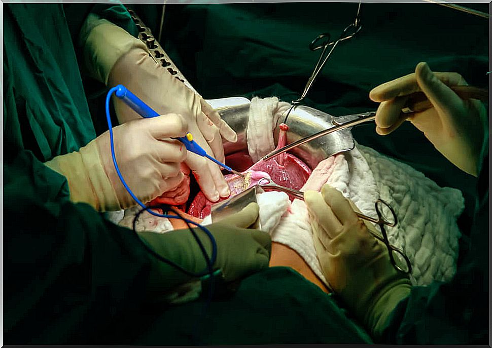
When the wound is located in the chest area, the usual treatment to follow consists of performing a posterolateral thoracotomy or a medial thoracotomy or sternotomy.
The surgical technique of posterolateral thoracotomy basically includes the following procedures: general anesthesia, sectioning of the muscle with the electrosurgical unit, opening of the pleura and detection of the vascular lesion.
The surgical technique of medial thoracotomy or sternotomy includes : general anesthesia, opening of the sternum with an electric saw, observation and placement of a forceps or clamp; location of the vascular lesion and realization of the graft.
In both cases, at the end it must be verified that the procedure has been done correctly and that there are no leaks. Subsequently , a drainage medium is created, the wound is closed, hemostasis is checked, the skin is sutured, and a dressing is placed.
Treatment of the great abdominal vessels
If the wound is abdominal, the usual thing is to go to the laparotomy. In this, first of all general anesthesia is applied. The operative area is then exposed, using devices called Bader Farebeuf retractors. These are operated manually.
After this step, the abdominal fascia is sectioned. This is a very resistant membranous structure. Then the disclosure of the rectus muscles is carried out. This is a maneuver to separate the tissues, without injuring them. It is done without a sharp instrument.
Immediately the spacers are rearranged, the peritoneum is divided, the bleeding vessel is identified and it is corrected. In this way, the prosthesis or graft is placed and the cavity is washed if necessary.
As in the other procedures, here homeostasis is also checked and it is verified that there are no leaks, a means of drainage is left or the so-called “Bogotá bag” is put on. Then, if possible, it is closed and sutured.