What Types Of Granulomas Are There?
There are various types of granulomas that manifest as a response of the body to an aggressive agent that cannot be resolved in the usual way. That is, we are facing an injury that denotes the inability of the defense cells to carry out an adequate elimination.
By definition, a granuloma is an accumulation of cells in the form of a tumor around something that is identified as foreign to the body. The cells in question belong to the immune system and the foreign agent can be a microorganism or an inert particle.
The above is what will determine the different clinical pictures, although similar if seen under a microscope. To learn more about it, below we detail the characteristics of each one.
Types of granulomas
Once a granuloma has started, the types that can become present depend on what you are trying to remove. At the same time, the varieties differ in the product of the response and in the possibility of being limited in one area or expanding to more tissues in an uncontrolled way.
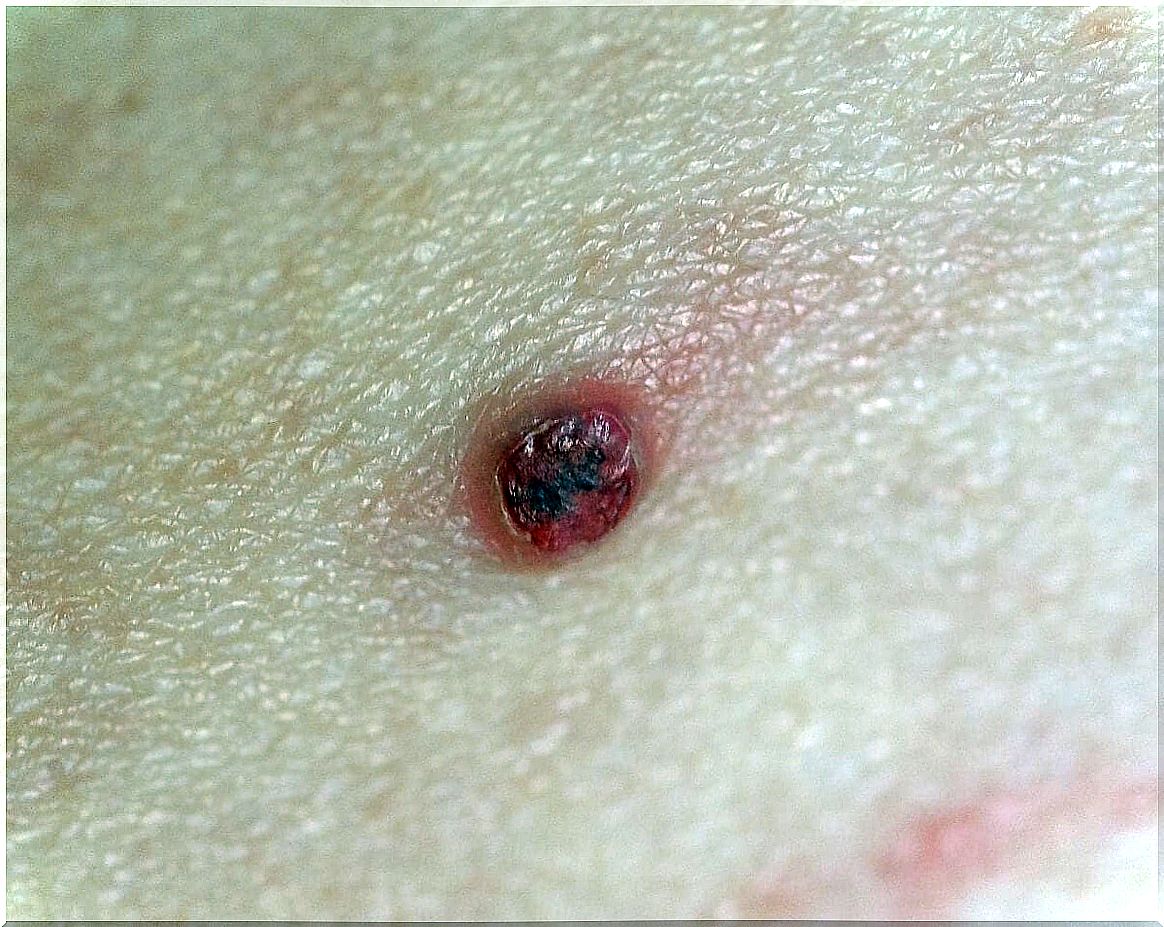
Pyogenic granuloma
One of the types of granulomas is pyogenic. Although there are no specific causes, it is associated with microorganisms that enter under the skin through small wounds, and that are encapsulated by the immune system without completely eliminating it to the outside.
The process takes time, since the injury occurs first and, later, the granulomatous reaction. The most frequent sites of appearance are the upper extremities and the face. This is assumed to be the case due to the higher frequency of superficial wounds that usually occur in these anatomical areas.
The clinical form is a lump, like a benign tumor. They tend to have repetitive bleeding that occurs by rubbing, scratching or spontaneously. The red color with which they are detected and seen is due to the number of small blood vessels they have.
Many granulomas go away without treatment. The explanation would be that the body, finally, manages to resolve the expulsion of the foreign body present. In other cases, it is necessary to intervene, either with incisions or laser surgery to remove them.
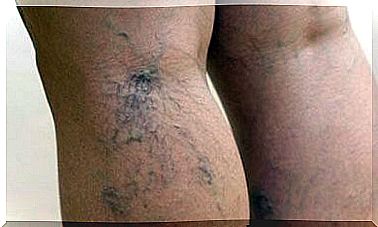
Granuloma annulare
As the name implies, these types of granulomas have a characteristic ring-shaped lesion. The skin protrudes in reddish circles, especially on the hands and feet.
The most common way is localized. In these cases, there are injurious rings, of about 5 centimeters, that are concentrated in specific anatomical regions, without expanding to the rest of the body.
The cause is considered unknown, although risk factors that favor its appearance have been identified. Diabetic patients, for example, are more susceptible. Cancer people are also affected by a repetitive variety of granuloma annulare that is difficult to treat.
Symptoms are mild, painless. Sometimes there is an almost imperceptible itch. As in pyogenic granuloma, the evolution can lead to spontaneous resolution or require an interventional approach.
Generalized granuloma annulare
The generalized form of granuloma annulare differs from the previous one in the expansion that occurs between anatomical areas of the body. The lesions are ring-shaped, reddish, with slight itching, almost all over the skin.
It can be resolved spontaneously, but it usually takes 2 years to complete the entire cycle from its appearance. Its priority population is adult women, especially those over 40 years of age. It does not become malignant, despite its extent. Nor can it be linked to a specific infection.
The most effective treatment is with a combination of antibiotics. The most widely used regimen uses rifampicin, ofloxacin, and minocycline. Sometimes corticosteroids and even immunomodulators are prescribed to help.
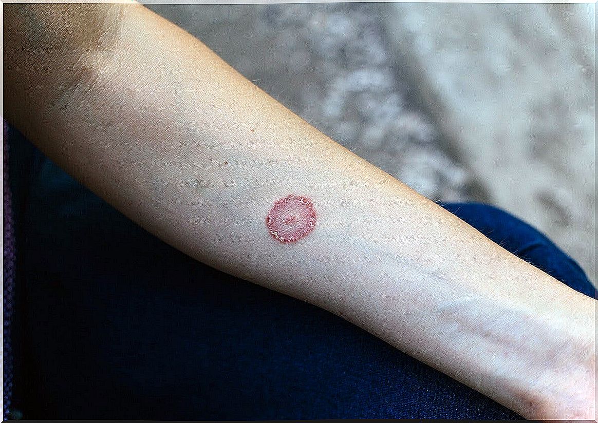
Perforating granuloma annulare
As another type of granuloma, a variety of the previous annulars, we have the perforator. It is also known as perforans and the etiology is not defined. Insect bites, UV exposure, hormonal imbalances from insulin, and hypothyroidism have been speculated on.
It can be accompanied by vasculitis near dermal lesions. This is an inflammation of the blood vessels that break more easily, heal poorly and are expressed in subcutaneous hemorrhages such as punctates or petechiae.
The characteristic sign of the perforating granuloma is its navel appearance. In the center of the annulus of the lesion there is usually a collapse of the tissue that is covered with a viscous substance. This fluid is collagen that seeps from under the skin to the surface. They can be on the hands, abdomen, and back.
The treatment is not so consensual due to how unusual the pathology is. Corticosteroids tend to be combined with antihistamines, as well as immunomodulators. The drugs are prescribed orally or locally (creams). If necessary, laser or phototherapy is applied.
The diagnosis of each type of granuloma is the beginning
This pathology requires proper treatment, but more importantly, an accurate diagnosis. The types of granulomas require the intervening dermatologist to have an acute capacity for analysis and interpretation of biopsies.
Although it tends to be limited in time, it is a disorder that generates discomfort and for which it is frequently consulted to evaluate therapeutic options. It will always be preferable that a health professional weighs the alternatives and offers the most appropriate for the case.